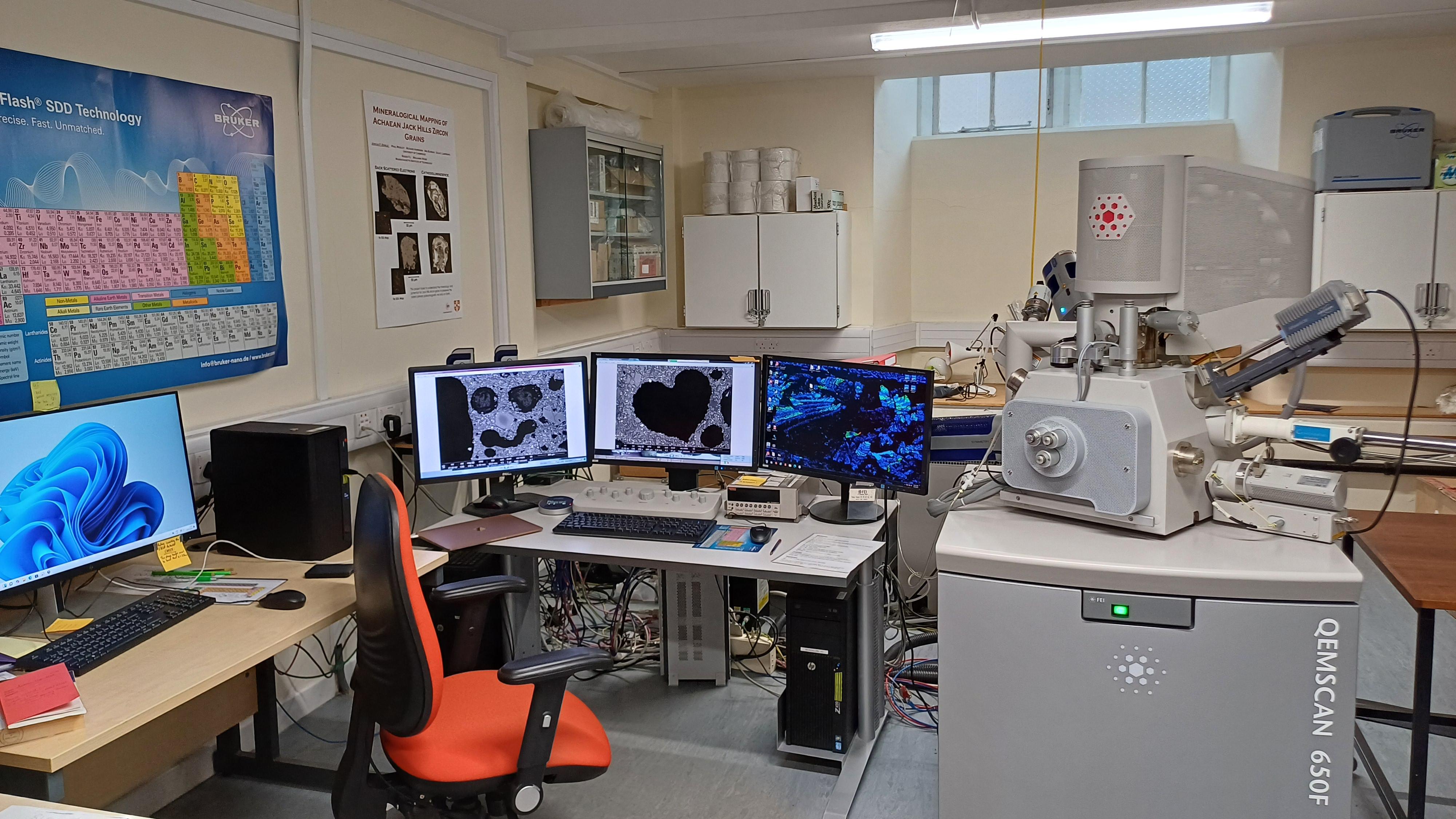
In October 2014, we took delivery of a Field Emission Gun Scanning Electron Microscope (FEG-SEM). The Quanta-650F is equipped with BSE, SE, GSED, LFD, panchromatic CL and in February 2024 was fitted with an Oxford Instruments Symmetry S3 EBSD and Ultim Max 170 EDS detectors. Several software packages including MAPS, Esprit (for Bruker EDS/EBSD data collected pre February 2024) and a full AZtec 6.2 licence with Mapsweeper and AZtecCrystal allow for a comprehensive list of techniques to be used in tandem to fully explore and interrogate a given sample.
1. Academic Staff Member: Dr Oscar Branson
2. Contact: Please email the lab managers or come find us in our offices.
Peak demand for the laboratory typically occurs from October to January. At other times of the year, the SEM is usually over-subscribed and waiting times are typically 2-5 weeks. Please do talk to us if you have an urgent requirement. To book a slot, please email Iris and Rosa.
______________________________________________________________________________________________
From 1st December 2024, please do the following:
To look at availability and book, please look at: PPMS
NOTE: to book a slot for this instrument you will need to have read and signed:
- All health and safety forms
- agree to the terms and conditions of the lab and computer rules
- book and complete training
Before being signed off as an independent user your session will have to be approved by Dr Iris Buisman and/or Dr Rosa Danisi. As an independent user you will be able to book as you please.
______________________________________________________________________________________________
Training videos for setting up the SEM: All users are requested to watch these online training videos before being granted access.
3. Quick overview of different techniques
The SEM is a fully equipped instrument that can be run at high vacuum, low vacuum or environmental mode. This allows for imaging/chemically analysing samples with or without a conductive (carbon and/or gold) coat.
Imaging
- Secondary Electron (SE) – A common imaging technique that collects low energy (<50 eV) secondary electrons that are ejected from the k-shell in the sample by inelastic scattering interactions with the incident beam electrons. They originate from within a few nanometres from the sample surface, thus providing an excellent, high resolution, depth of field image. This is typically used for imaging structures and topography of samples.
- Back Scattered Electrons (BSE) – These electrons vary directly as a function of composition by virtue of the atomic number (Z) through inelastic interactions in the specimen. Provides an invaluable imaging tool to rapidly discriminate phases that have different mean Z values.
- Cathodoluminescence (CL) – A signal produced upon the promotion of a valence band electron to the conduction band. The resulting photon emission of electromagnetic radiation is within the visible light region.
All three of these signals can be obtained of large areas of a given sample using a stage mapping software, MAPS.
Chemical/Phase Analysis
- Energy Dispersive X-Ray Spectroscopy (EDS) Spot Analysis and Mapping – Using characteristic X-rays for elemental analysis or chemical characterisation of a given sample. This is a relatively simple and easy technique for major and some minor elemental analysis.
- Electron Back Scattered Diffraction (EBSD) – Is a technique that uses electron diffraction signal to characterise the texture of polycrystalline material. Typically, it is used to explore texture at the millli- to micrometer scale, grain morphology and strain deformation.
Other features of the SEM allow for Beam deceleration mode, charge contrast imaging (CCI) and Environmental SEM (ESEM) mode options.
